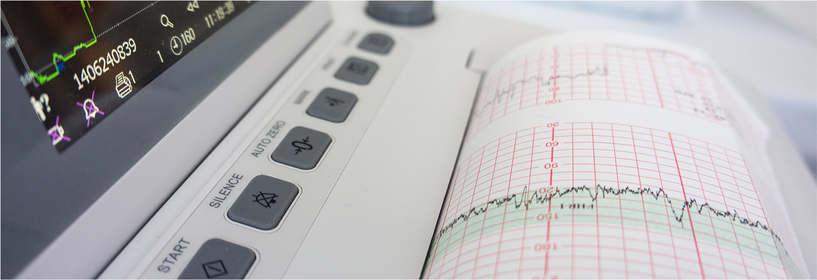
All Echocardiography (Echo) procedures involve an ultrasound machine that uses harmless sound waves to capture images and movies of the heart.
These images give the doctor information on the heart muscle, valves, blood vessels and blood flow. From these images, the size, structure and function of the heart can be measured and evaluated.
Patients may be referred for an Echo to investigate structural abnormalities, heart murmurs, an enlarged heart or to exclude structural heart disease. An Echo may also be performed to form a baseline for future investigations.
Echo procedures are all conducted using a specialised probe and gel placed at several locations on the chest. All procedures are performed while the patient is lying on their left side.
For more information on Echo procedures performed by our cardiologists, please select from the list below:
